Physics on the Nanometer Scale
SCIENCE  SCIENCE
| ...last update: January 2016 |
| Science |
| Teaching |
| People - Contact |
| Movies |
| STARTPAGE |
Our research is devoted to employing physics tools and concepts to understand and modify molecular objects with a main emphasis on biological systems |
LIST OF PROJECTS:
|
Exploring the Properties of Individual DNA Molecules in the liquid phase by video fluorescent microscopy. |
Holography with Low Energy Electrons is used to image individual DNA molecules and proteins. It also allows real time (with 30 images per second) observation of nucleation and growth as well as the study of charges and their transfer or transport in molecules. |
Preparation and Structural Determination of Individual Biomolecules aims at structural biology at the single molecule level. |
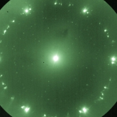
Coherent Low-Energy Electron Diffraction Microscopy is a project aiming at obtaining atomic resolution structural information from oversampled low energy electron diffraction patterns. It is supported by the Swiss National Science Foundation. Novel phase retrieval methods in coherent diffraction from non-crystalline objects of nanometer dimensions is a related project aiming at the numerical reconstruction of holograms and coherent diffraction patterns. The EU Project SIBMAR - with partners from Brno, Manchester and Zurich - has been completed and here is a short account - by the EU commission - about the achievements of SIBMAR.
|
|
RECENT PUBLICATIONS: 2017: Resolution enhancement in in-line holography by numerical compensation of vibrations, by Tatiana Latychevskaia and Hans-Werner Fink - Optics Express Vol. 25, Issue 17, pp. 20109-20124 (2017) Imaging the potential distribution of individual charged impurities on graphene by low-energy electron holography, by Tatiana Latychevskaia, Flavio Wicki, Conrad Escher, Hans-Werner Fink - Ultramicroscopy, Volume 182, November 2017, Pages 276-282 Imaging proteins at the single molecule level, by Jean-Nicolas Longchamp, Stephan Rauschenbach, Sabine Abb, Conrad Escher, Tatiana Latychevskaia, Klaus Kern, Hans-Werner Fink - PNAS, February 14, 2017, vol. 114 no. 7 2016: In relation to the publication "Practical algorithms for simulation and reconstruction of digital in-line holograms", Appl. Optics 54, 2424 - 2434 (2015), Tatiana Latychevskaia has published the corresponding MATLAB code here (October 2016) Direct observation of individual elementary charges and their dynamics on graphene by low-energy electron holography, byTatiana Latychevskaia, Flavio Wicki, Jean-Nicolas Longchamp, Conrad Escher, Hans-Werner Fink, Nano Letters, 2016, 16 (9), pp 5469–5474 Creating Airy beams employing a transmissive spatial light modulator, by Tatiana Latychevskaia1, Daniel Schachtler and Hans-Werner Fink, Applied Optics, Vol. 55, Issue 22,
pp. 6095-6101, (2016)
Inverted Gabor holography principle for tailoring arbitrary shaped three-dimensional beams, by Tatiana Latychevskaia & Hans-Werner Fink, Nature - Scientific Reports 6, Article number: 26312 (2016) Mapping Unoccupied Electronic States of Freestanding Graphene by Angle-Resolved Low-Energy Electron Transmission, by Flavio Wicki, Jean-Nicolas Longchamp, Tatiana Latychevskaia, Conrad Escher & Hans-Werner Fink, Phys. Rev. B 94, 075424 – Published 16 August 2016 Resolution enhancement by extrapolation of coherent diffraction images: a quantitative study on the limits and a numerical study of nonbinary and phase objects, by T. Latychevskaia, Y. Chushkin & H.-W. Fink, Journal of Microscopy, 2016, 264: 3–13. doi:10.1111/jmi.12408
Design and Implementation of a Micron-Sized Electron Column Fabricated by Focused Ion Beam Milling, by Flavio Wicki, Jean-Nicolas Longchamp, ConradEscher and Hans-Werner Fink, Ultramicroscopy, Volume 160, January 2016, Pages 74–79 2015: Imaging outside the box: Resolution enhancement in X-ray coherent diffraction imaging by extrapolation of diffraction patterns, by Tatiana Latychevskaia, Yuriy Chushkin, Federico Zontone and Hans-Werner Fink, Appl. Phys. Lett. 107, 183102 (2015) Low-energy electron holographic imaging of individual tobacco mosaic virions, by Jean-Nicolas Longchamp, Tatiana Latychevskaia, Conrad Escher and Hans-Werner Fink, Appl. Phys. Lett. 107, 133101 (2015) Holography and Coherent Diffraction with Low-Energy Electrons: A Route towards Structural Biology at the Single Molecule Level, by Tatiana Latychevkaia, Jean-Nicolas Longchamp, Conrad Escher, Hans-Werner Fink, Ultramicroscopy - online version Reconstruction of purely absorbing, absorbing and phase-shifting, and strong phase-shifting objects from their single-shot in-line holograms by Tatiana Latychevskaia and Hans-Werner Fink, Applied Optics, Vol. 54, Issue 13, pp. 3925-3932 (2015) Atomically resolved structural determination of graphene and its point defects via extrapolation assisted phase retrieval, by Tatiana Latychevskaia and Hans-Werner Fink, Appl. Phys. Lett. 106, 021908 (2015) Practical algorithms for simulation and reconstruction of digital 2014: Interfacing Ultraclean Graphene with Solid-State Devices, by Jean-Nicolas Longchamp, Conrad Escher, Hans-Werner Fink, preprint Holographic time-resolved particle tracking by means of three-dimensional volumetric deconvolution by Tatiana Latychevskaia and Hans-Werner Fink Optics Express, Vol. 22, Issue 17, pp. 20994-21003 (2014), http://dx.doi.org/10.1364/OE.22.020994 Atomically resolved structural determination of graphene and its point defects by employing extrapolation assisted phase retrieval by Tatiana Latychevskaia and Hans-Werner Fink, preprint Resolution enhancement in digital holography by self-extrapolation of holograms by Tatiana Latychevskaia and Hans-Werner Fink, in Advances in Engineering / Applied Physics On artefact-free reconstruction of low-energy (30-250 eV) electron holograms, by Tatiana Latychevskaia, Jean-Nicolas Longchamp, Conrad Escher, Hans-Werner Fink, submitted to a special edition of Ultamicroscopy devoted to Low Voltage Electron Microscopy; in press http://www.sciencedirect.com/science/article/pii/S0304399113003124 Low-energy electron holographic imaging of ultraclean graphene-supported gold-nanorods, by Jean-Nicolas Longchamp, Conrad Escher, Tatiana Latychevskaia and Hans-Werner Fink, submitted to a special edition of Ultamicroscopy devoted to Low Voltage Electron Microscopy; in press http://www.sciencedirect.com/science/article/pii/S0304399114000102 2013: Coherent microscopy at resolution beyond diffraction limit using post-experimental data extrapolation, by Tatiana Latychevskaia and Hans-Werner Fink, Appl. Phys. Lett. 103, 204105 (2013); http://dx.doi.org/10.1063/1.4831985 Graphene Unit Cell Imaging by Holographic Coherent Diffraction, by Jean-Nicolas Longchamp, Tatiana Latychevskaia, Conrad Escher, Hans-Werner Phys. Rev. Lett. 110, 255501 (2013) http://prl.aps.org/abstract/PRL/v110/i25/e255501 Pulsed Electron Holography, by Matthias Germann, Tatiana Latychevskaia, Conrad Escher, and Hans-Werner Fink, Appl. Phys. Lett. 102, 203115 (2013) http://dx.doi.org/10.1063/1.4807661 Resolution enhancement in digital holography by self-extrapolation of holograms by Tatiana Latychevskaia and Hans-Werner Fink, Optics Express, Vol. 21, Issue 6, pp. 7726-7733 (2013) Coherent Diffraction and Holographic Imaging of Individual Biomolecules Using Low-Energy Electrons by Tatiana Latychevskaia, Jean-Nicolas Longchamp, Conrad Escher, and Hans-Werner Fink, in Advancing Methods for Biomolecular Crystallography, Springer Verlag (2013), DOI: 10.1007/978-94-007-6232-9_29 Ultra-Clean Freestanding Graphene by Platinum-Metal Catalysis by Jean-Nicolas Longchamp, Conrad Escher, Hans-Werner Fink, J. Vac. Sci. Technol. B 31, 020605 (2013) 2012: When holography meets coherent diffraction imaging by Tatiana Latychevskaia, Jean-Nicolas Longchamp and Hans-Werner Fink,
Optics Express 20(27), pp. 28871-28892 (2012) Fabrication of metallic double-gate field emitter arrays and their electron beam collimation characteristics by Partick Helfenstein, Konstantin Jefimovs, Eugenie Kirk, Conrad Escher, Hans-Werner Fink and Soichiro Tsujino Ultra-Clean Freestanding Graphene by Platinum-Metal Catalysis by Jean-Nicolas Longchamp, Conrad Escher, Hans-Werner Fink, http://arxiv.org/abs/1210.6824 Low-energy electron transmission imaging of clusters on free-standing graphene by Jean-Nicolas Longchamp, Tatiana Latychevskaia, Conrad Escher, Hans-Werner Fink, Appl. Phys. Lett. 101, 113117 (2012); doi: 10.1063/1.4752717 Non-destructive imaging of an individual protein by J.-N. Longchamp, T. Latychevskaia, C. Escher, and H.-W. Fink, Appl. Phys. Lett. 101, 093701 (2012); http://dx.doi.org/10.1063/1.4748113 |
2011: Novel Fourier-domain constraint for fast phase Individual filamentous phage imaged by electron holography, Gregory B. Stevens, Michael Krüger, Tatiana Latychevskaia, Peter Lindner, Andreas Plückthun, Hans-Werner Fink, European Biophysics Journal, Biophysics Letters, doi: 10.1007/s00249-011-0743-y (2011) Highly collimated electron beams from double-gate field emitter arrays with large collimation gate apertures, P. Helfenstein, E. Kirk, K. Jefimovs, T. Vogel, C. Escher, H.-W. Fink, and S. Tsujino, Appl. Phys. Lett. 98, 061502 (2011); doi:10.1063/1.3551541 Coherent low-energy electron diffraction on individual nanometer sized objects, E. Steinwand, J.-N. Longchamp, H.-W. Fink, Ultramicroscopy 111(4), pp. 282-284 (2011); doi:10.1016/j.ultramic.2010.12.018 Fabrication and characterization of low aberration micrometer-sized electron lenses, E. Steinwand, J.-N. Longchamp, H.-W. Fink, Ultramicroscopy 110(9), pp.1148-1153 (2010); doi:10.1016/j.ultramic.2010.04.013 Field-Emission Characteristics of Molded Molybdenum Nanotip Arrays With Stacked Collimation Gate Electrodes, S. Tsujino, P. Helfenstein, E. Kirk, T. Vogel, C. Escher, H.-W. Fink, IEEE Electron Device Letters 31(9), pp.1059-1061 (2010); 10.1109/LED.2010.2052013 Off-axis and inline electron holography: Experimental comparison, Tatiana Latychevskaia, Petr Formanek, C. T. Koch and Axel Lubk, Ultramicroscopy 110(5), pp. 472-482 (2010);doi:10.1016/j.ultramic.2009.12.007 Depth-resolved holographic reconstructions by three-dimensional deconvolution Non-destructive Imaging of Individual Bio-Molecules, Matthias Germann, Tatiana Latychevskaia, Conrad Escher & Hans-Werner Fink, PDF-File: Phys. Rev. Lett. 104, 095501 (2010).....PRL-Webseite.....Movie about the Experiment doi:10.1103/PhysRevLett.104.095501 2009: Simultaneous reconstruction of phase and amplitude contrast from a single holographic record, Tatiana Latychevskaia and Hans-Werner Fink, Optics Express, 17(13), pp. 10697-10705 (2009); doi:10.1364/OE.17.010697 Solution of the Twin Image Problem in Holography, Tatiana Latychevskaia and Hans-Werner Fink, Phys. Rev. Lett. 98, 233901 (2007); doi:10.1103/PhysRevLett.98.233901 Zupfen am Lebensfaden: Experimente mit einzelnen DNA Molekülen, Conrad Escher und Hans-Werner Fink, “Physik in unserer Zeit”, Wiley-VCH Verlag , Heft 4, 2007 2006: A Quantum Mechanical Scheme to reduce Radiation Damage in Electron Microscopy, Hiroshi Okamoto, Tatiana Latychevskaia and Hans-Werner Fink, doi:10.1063/1.2191096, Appl. Phys. Lett. 88, 164103 (2006); doi:10.1063/1.2191096 Cryogenic Low Energy Electron Point Source Microscope, Hiroshi Okamoto and Hans-Werner Fink, doi:10.1063/1.2195120, Rev. Sci. Instrum. 77, 043714 (2006); doi:10.1063/1.2195120 Vacuum Ion Emission from Solid Electrolytes, Conrad Escher, Sandra Thomann, Cornel Andreoli, Hans-Werner Fink, Julien Toquant and Dieter Pohl, Appl. Phys. Lett. 89, 053513 (2006); doi:10.1063/1.2264092 Direct Evidence for Conduction Pathways in Solid Electrolytes, Conrad Escher, Tatiana Latychevskaia, Hans-Werner Fink and Dieter Pohl, Phys. Rev. Lett. 97, 136601 (2006); doi:10.1103/PhysRevLett.97.136601 Patents:
Popular science reading (in German):
|
 Annual Report - April 2016 to April 2017
Summary of selected recent achievements |
|
Imaging proteins at the single molecule level, by Jean-Nicolas Longchamp, Stephan Rauschenbach, Sabine Abb, Conrad Escher, Tatiana Latychevskaia, Klaus Kern, Hans-Werner Fink - PNAS - read more
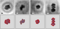
PNAS, February 14, 2017, vol. 114 no. 7 All established methods for obtaining structural information about proteins require averaging over thousands or even millions of molecules, be it due to radiation damage or a too weak signal from just one individual protein. Since proteins are flexible objects that can assume distinct conformations often associated with different functions, averaging over a large ensemble of proteins leaves these associated structural details undiscovered. +++++++++++++++++++++++ CHARGE TRANSPORT MEASUREMENTS ON GRAPHENE
This thesis investigates the electronic properties of graphene rendered ultraclean by a palladium-metal catalysis. Additionally Raman spectroscopy is carried out to further investigate the properties of the ultraclean graphene. The research objectives can be summarized in the following way: +++++++++++++++++++++++ Low-energy electron holographic imaging of individual tobacco mosaic virions by Jean-Nicolas Longchamp, Tatiana Latychevskaia, Conrad Escher and Hans-Werner Fink - read more
Modern structural biology relies on Nuclear Magnetic Resonance (NMR), X-ray crystallography, and cryo-electron microscopy for gaining information on biomolecules at nanometer, sub-nanometer, or atomic resolution. All these methods, however, require averaging over a vast ensemble of entities, and hence knowledge on the conformational landscape of an individual particle is lost. Unfortunately, there are now strong indications that even X-ray free electron lasers will not be able to image individual molecules but will require nanocrystal samples. Here, we show that non-destructive structural biology of single particles has now become possible by means of low-energy electron holography. As an example, individual tobacco mosaic virions deposited on ultraclean freestanding graphene are imaged at 1 nm resolution revealing structural details arising from the helical arrangement of the outer protein shell of the virus. Since low-energy electron holography is a lens-less technique and since electrons with a deBroglie wavelength of approximately 1 Å do not impose radiation damage to biomolecules, the method has the potential for Angstrom resolution imaging of single biomolecules. In relation to the above article: Milestone single-biomolecule imaging technique may advance drug design by Zhengzheng Zhang, the American Institute of Physics, an AIP publication Atomically resolved structural determination of graphene and its point defects by employing extrapolation assisted phase retrieval by Tatiana Latychevskaia and Hans-Werner Fink, Appl. Phys. Lett. 106, 021908 (2015) - read more We demonstrate that the structure of a nanocrystal can unambiguously be recovered from its diffraction pattern alone by applying a direct phase retrieval procedure not relying on prior information of the object shape. Individual point defects in the atomic lattice are clearly apparent. We summarize the conditions for this phase retrieval method and show that the diffraction pattern can be extrapolated beyond the original record to even reveal formerly not visible Bragg peaks. Such extrapolated wave field pattern leads to enhanced spatial resolution in the reconstruction. Coherent microscopy at resolution beyond diffraction limit using post-experimental data extrapolation, by Tatiana Latychevskaia and Hans-Werner Fink, Appl. Phys. Lett. 103, 204105 (2013) read more Conventional microscopic records represent intensity distributions whereby local sample information is mapped onto local information at the detector. In coherent microscopy, the superposition principle of waves holds; field amplitudes are added, not intensities. This non-local representation is spread out in space and interference information combined with wave continuity allows extrapolation beyond the actual detected data. Established resolution criteria are thus circumvented and hidden object details can retrospectively be recovered from just a fraction of an interference pattern. Graphene Unit Cell Imaging by Holographic Coherent Diffraction, by Jean-Nicolas Longchamp, Tatiana Latychevskaia, Conrad Escher, Hans-Werner Phys. Rev. Lett. 110, 255501 (2013) read more We have imaged a freestanding graphene sheet of 210 nm in diameter with 2 Å resolution by combining coherent diffraction and holography with low-energy electrons. The entire sheet is reconstructed from a single diffraction pattern displaying the arrangement of 660.000 individual graphene unit cells at once. Given the fact that electrons with kinetic energies of the order of 100 eV do not damage biological molecules, it will now be a matter of developing methods for depositing individual proteins onto such graphene sheets. Low-energy electron transmission imaging of clusters on free-standing graphene by Jean-Nicolas Longchamp, Tatiana Latychevskaia, Conrad Escher, Hans-Werner Fink, read more We investigated the utility of free-standing graphene as a transparent sample carrier for imaging nanometer-sized objects by means of low-energy electron holography. The sample preparation for obtaining contamination-free graphene as well as the experimental setup and findings are discussed. For incoming electrons with 66 eV kinetic energy graphene exhibits 27% opacity per layer. Hence, electron holograms of nanometer-sized objects adsorbed on free-standing graphene can be recorded and numerically reconstructed to reveal the object’s shapes and distribution. Furthermore, a Moiré effect has been observed with free-standing graphene multi-layers. Non-destructive imaging of an individual protein by J.-N. Longchamp, T. Latychevskaia, C. Escher, and H.-W. Fink, read more Imaging a single biomolecule at atomic resolution without averaging over different conformations is the ultimate goal in structural biology. We report recordings of a protein at nanometer resolution obtained from one individual ferritin by means of low-energy electron holography. One single protein could be imaged for an extended period of time without any sign of radiation damage. Since the fragile protein shell encloses a robust iron cluster, the holographic reconstructions could also be cross-validated against transmission electron microscopy images of the very same molecule by imaging its iron core. When holography meets coherent diffraction imaging. We discovered a mathematical relationship between a hologram and a diffraction pattern allowing us to merge the two prominent phase retrieval methods into one leading to fast and unambiguous object reconstructions while not requiring the oversampling condition anymore. read more Coherent low-energy electron diffraction of individual nanometer sized objects. The first coherent diffraction pattern of an individual free-standing carbon nanotube using low-energy electrons has been obtained. read more Fabrication and characterization of low aberration micrometre-sized electron lenses. We have developed methods for the routine fabrication of micron sized electron lenses and evaluated their performance in a dedicated UHV vacuum system. A detailed evaluation of the spherical aberrations shows that these lenses have a quality that can otherwise only be reached by elaborate magnetic objective lenses in modern electron microscopes. read more |
|
Pulsed electron holography has been invented to circumvent the disturbing effects of residual vibrations, drift and ac-magnetic fields limiting the coherence of the low energy electron wave. It has been possible to acquire a hologram of a single DNA within 10 micro-seconds. A set of several 100 such holograms of one and the same molecule is used to build up a record with a high signal to noise level. For this, the maximum of the cross correlation between subsequently acquired holograms is computed prior to shifting and superimposing the whole set of pulsed holograms. Ref.: Master thesis of Matthias Germann |
|
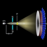
Quantitative Radiation Damage studies in Low Energy Electron Point Source Microscopy. While it seemed qualitatively apparent for quite some time that electron waves, associated with kinetic energies below 300eV, introduce little respectively non-detectable radiation damage on such fragile biological objects like an individual DNA molecule, a quantitative study of radiation damage has only recently been attempted. A set of holograms of a freshly prepared DNA sample has been acquired and the cross correlation between subsequent taken records been computed. Thereafter, the DNA molecule has been subject to continues radiation for one hour corresponding to a total effective dose of several orders of magnitude larger than what is considered tolerable in high energy TEM studies of biological samples. It should be noted that similar experiments have been carried out at slightly higher energies, namely above 300 eV. Under these conditions, a single DNA decomposes within a few 10 seconds of observation time. Future experiments should reveal if there is a well defined threshold for damage or if there exist discrete windows between a few 10 and a few 100 eV. Ref.: Master thesis of Matthias Germann Phys. Rev. Lett. 104, 095501 (2010) |
|
Solution of the Twin Image Problem in Holography. The reconstruction of a hologram is the final step that leads to the three dimensional shape of the scattered object wave that eventually reveals the structural detail of the object. While the quality of the reconstructed image depends on the quality of detection of amplitude and phase of the object wave in the holographic record, one intrinsic problem remains. It is the so-called “twin image problem” recognized since the invention of holography by Dennis Gabor. It is a consequence of the foundation of coherent optics that the out of focus conjugated twin is entangled with the wanted image and obscures its recognition. This long standing problem of coherent optics has recently been solved and provides object reconstructions no longer obscured by the twin image. Ref.: Phys. Rev. Lett. 98, 233901 (2007)
June 8, 2007: Molecular holograms are coming into focus: New Scientist comments on the twin image removal. June 19, 2007: Getting rid of the twin image that plagues holography: PhysOrg comments on the twin image removal. |
|
Energetics of a single DNA Molecule: Exploring the dynamics of individual DNA molecules has been subject of various theoretical studies and models over many decades, but only during the last 10 years the ability to actually carry out experiments on individual molecules has become routine. However, direct insight into the energetics on a single molecule level has not been obtained so far. We have carried out temperature dependent experiments with individual lambda-DNA molecules and provide the first quantitative information about the internal energetics of a single molecule in solution. From the mean square displacement of the center of mass of a DNA random coil we derive the diffusion coefficient from Einstein equation and from its temperature dependence the activation energy for diffusion to be roughly 1/6 eV. Following these control experiment we determine the activation energy associated with the transition from the straightened to the lowest free energy random coil configuration. The statistics of a total of 600 measurements on individual lambda-DNA molecules at 12 different temperatures reveals an activation free energy for the transition between these two prominent configurations of 1/4 eV. This indicates that the DNA random coil equilibrium configuration is not entirely given by entropy in contrast to current models. |
|
|
|
|
|
|