Forces in the Drosophila wing disc
Measuring the local stresses in wing imaginal discs
We employ a range of techniques, both optical and mechanical to determine the local stress distribution in tissues such as the wing imaginal disc. In order to determine the stresses directly, we employ photoelasticity directly on the tissue, where we are measuring the spatial distribution of birefringence to high accuracy. An example of the stress in a Drosophila wing imaginal disc using a birefringence setup is given in the figure below. As a control, stress was relieved by stretching the disc, clearly showing the stress nature of the birefringence signal.
When a disc grows, the stresses measured by photoelasticity increase with size, as would be expected from models of mechanical feedback of growth control. This can be seen in the figure below, where the size dependence of the birefringence in the centre as well as at the periphery is plotted. These latter data indicate that the stretching at the periphery remains constant as predicted by the model of T. Aegerter-Wilmsen et al. More details about this project can be found in our 2009 Mechanisms of Development paper in the publications section.
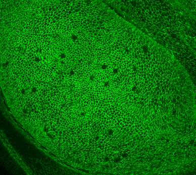
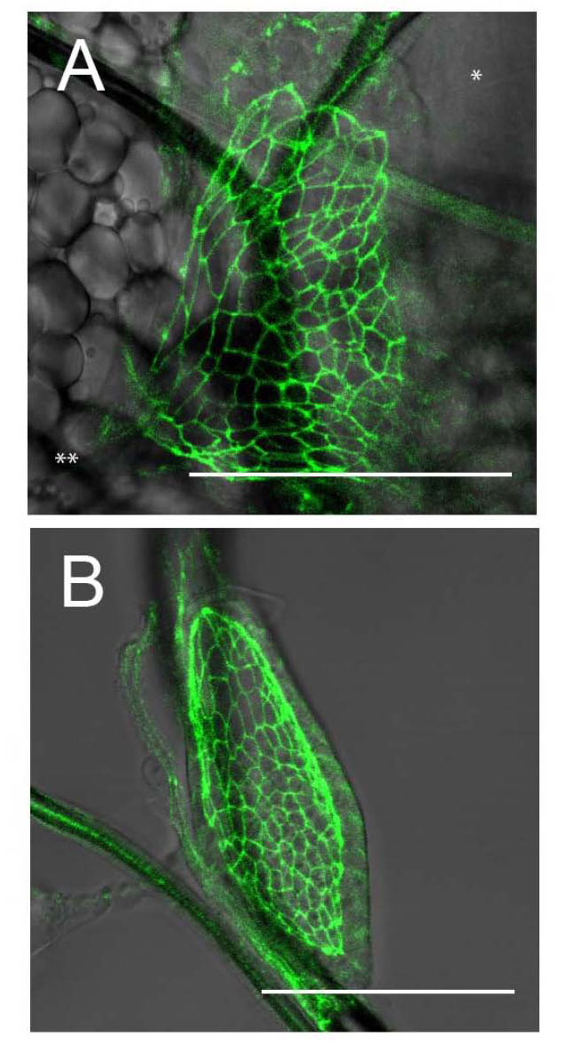
In order to study the (visco)elastic properties of the tissue, the strain also needs to be characterized, which we do by a quantification of cell shapes in the apical surface. An example of fluorescently marked cell shapes on the apical surface of a wing disc is shown below.
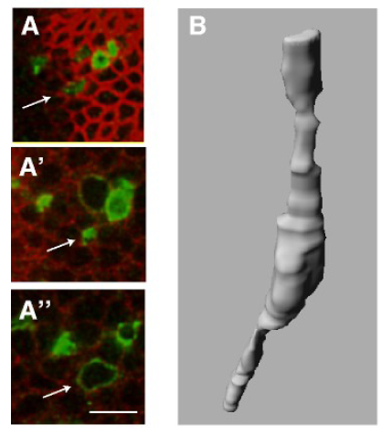
Apart from the apical cell shapes, which can be described by regular polygons, we have also characterized the three dimensional shape of single cells using mono-clonal analysis. The cell shapes outside the apical region are quite irregular and there is seems to be no correlation between the cell volume and the apical surface area.
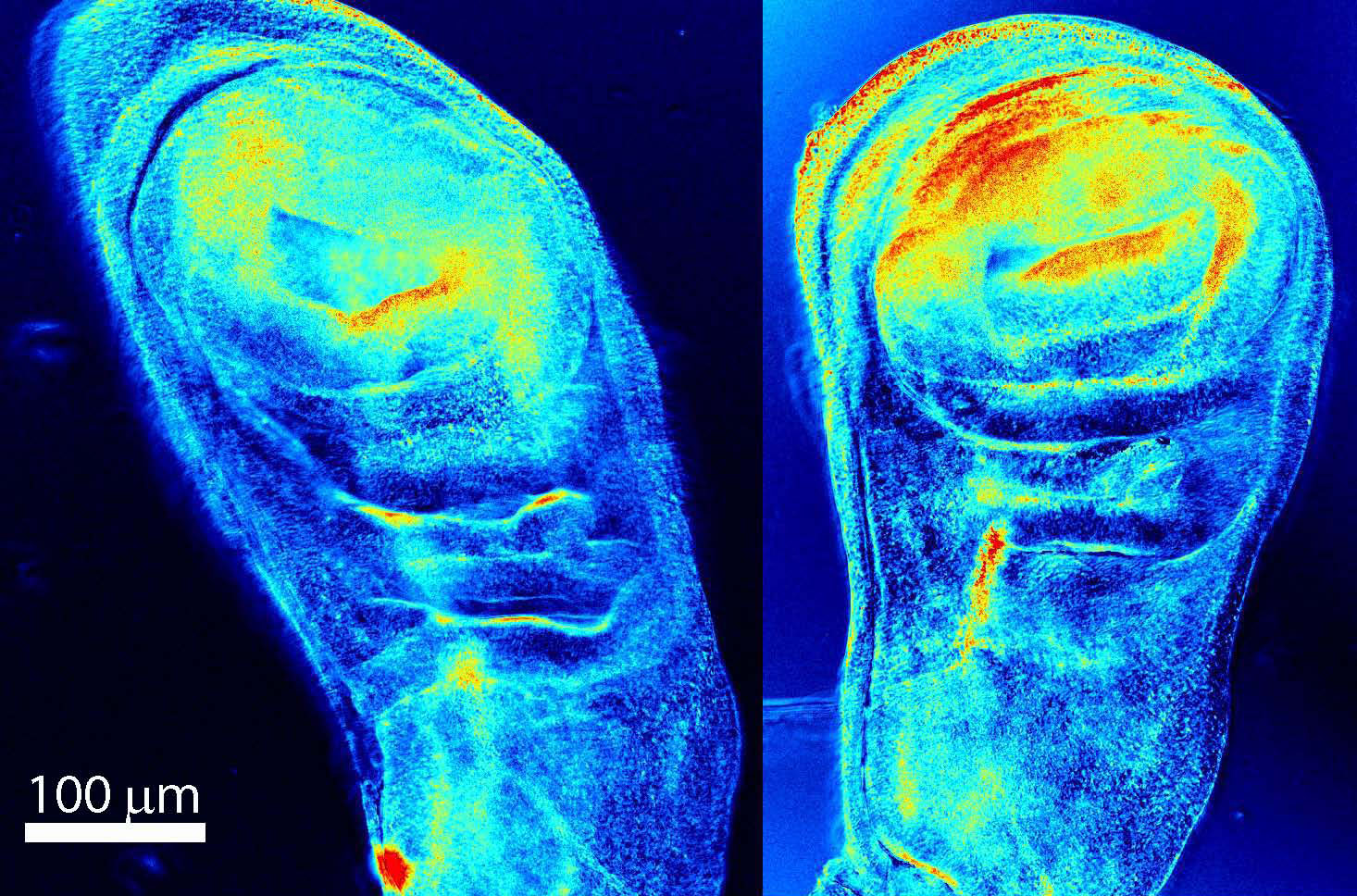
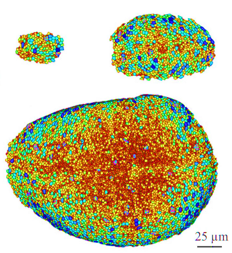
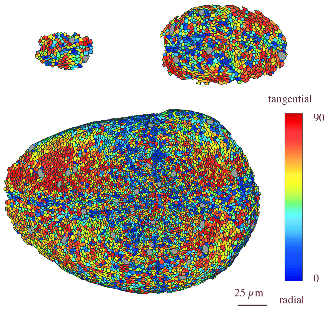
The apical cell shapes can now be used as an alternative measure of the development of mechanical forces acting on the cells during development. This is shown in the two figures below, where three different disc of different age ranging from the early to late third instar stages are shown. Here, apical cell outline markers have been used to study the strain of the cells. The apical area is quatified in the top figure, where red corresponds to small area, i.e. high compression. The bottom figure shows the elongation direction of the cell, where red shows tangential orientation. The to figures combined show that cells are compressed in the centre and tangentially stretched in the periphery, as predicted by the model discussed in the section about biological modelling. For more information consult our 2012 Development publication.
The genetic manipulations in terms of markers and the dissection of mechanosensation in Drosophila are done in collaboration with the Basler Lab at the University of Zurich.
Mechanical forces acting within the larva
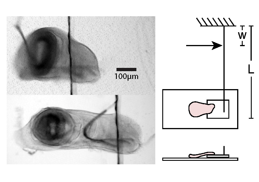
Using the method for in-vivo imaging described in the imaging section, we have studied the forces acting on wing discs within live larvae. This has shown that in the middle of the second instar stage, there are significant forces pulling on the wing disc. This is shown in the figures below. The top figure shows the disc in the larva, whereas the bottom figure shows the same disc right after dissecton. The scale bar is 50 microns. This shows the importance of mechanical forces in the development of the wing disc tissue. For more information consult the 2012 PLoSOne publication.
Direct mechanical stimulation of tissues
We are developing tools with which to pull on wing disc tissues with controlled forces in order to determine the influence of mechanical stresses on growth. Using such calibrated stimulation and the concurrent observation of the stress distribution, we are determining the spatial distribution of elastic properties as well as calibrating quantitatively the stress-optical parameters of the tissue at hand. With our setup, we are able to exert forces of the order of a few Micronewtons, which is in the relevant range for tissues and not commonly reachable by commercial techniques.
A wing disc before and after external stretching. By attaching the disc to two different cover slides, it is possible to exert a controlled force between a few Micronewton to a few Millinewton on the tissue. This allows a characterisation of the elastic as well as the stress-optical properties of the tissue. We obtain a photoelastic constant of the order of 2 10-10 Pa-1 for the wing imaginal disc. For more information, consult the 2010 EPJE in the publications section.
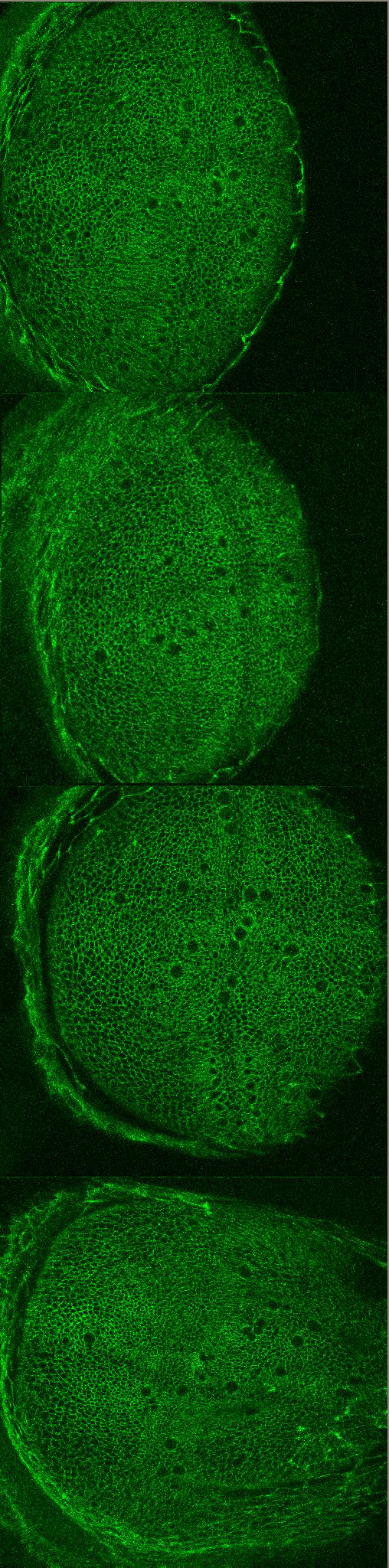
Using the above stretching apparatus, we have investigated the effect of tensional stresses on the proliferation rate of wing imaginal discs in the late third instar stage. Due to the fact that dividing cells are easily identifyable from their large and round shape in the apical side of the tissue, the proliferation rate of the tissue under mechanical tension can be determined. Representative images are shown below. On the top, an unstretched initial wing disc is shown. A similarly unstretched disc is shown after 1 hour in the next figure. The number of dividing cell has not changed significalntly. The third image shows a disc after 1 hour of strtching with a force of 160 Micronewton and similarly, the bottom figure a disc after stretching with 350 Micronewton for 1 hour. In both of these cases, there is a significant increase in the number of dividing cells.
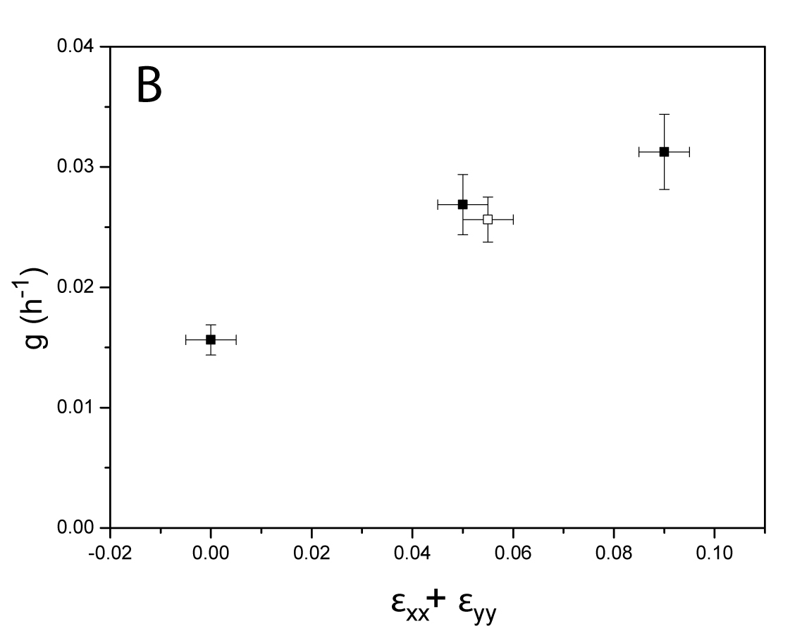
These data are quantified in the figure below, where the proliferation rate of the tissue is shown as a function of the applied total strain in the tissue. The open symobl corresponds to experiments, where the tissue was compressed, such that it buckled, leading to a different distribution of strain in the parallel and perpendicular direction. However, a tensional experiment with the same total strain leads to the same proliferation rate showing that the total strain is important in its determination. For more details, consult the 2013 PLoS One paper in the publications section.
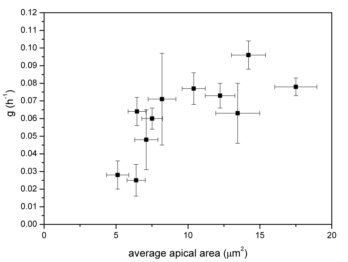
In order to study the effect of mechanical tension on proliferation, we have used in-vivo imaging to study the correlation between apical cell area, which is directly connected to the stress acting on the cells and the proliferation rate during the entire growth phase. The result is shown in the figure below, where a clear correlation can be seen.