In-vivo imaging
In-vivo Imaging of Drosophila larvae
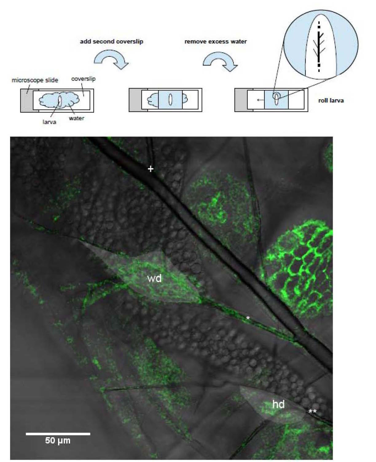
Using a combination of larval positioning and index matching of the cuticule we have developed a method with which to image larval development over the whole period of development. The basic method of the positioning is shown in the figure below. The wing disc is located with the help of where it is attached to the dorsal main trachea.
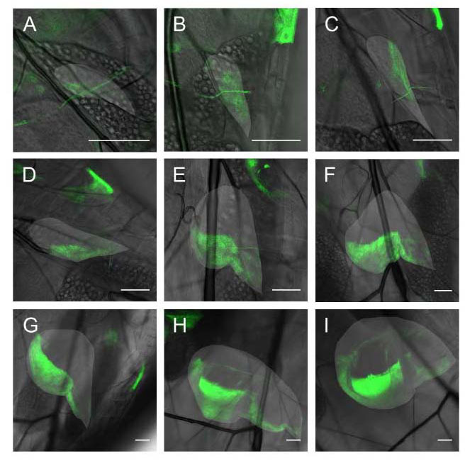
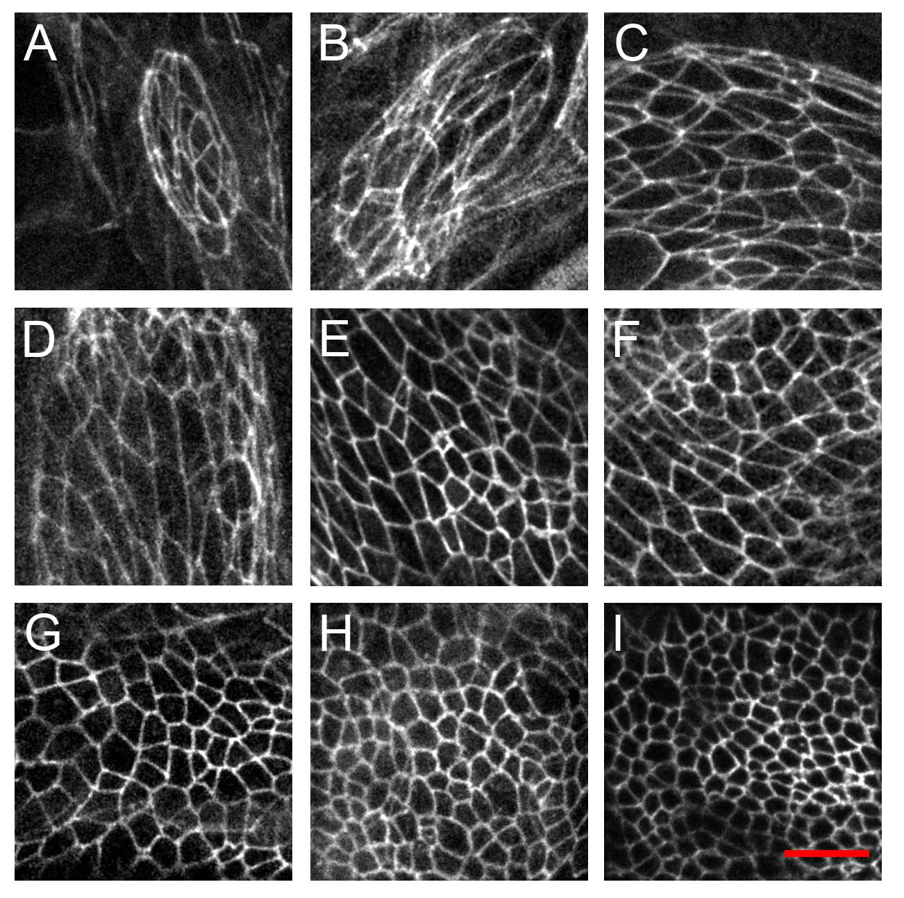
With this method it is possible to image larval structures, such as the wing disc at many time point during development with a time resolution of 4-8 hours. As an example of this, below we show the pattern of engrailed expression from the first instar to the third instar (scale bar 50 microns in all images) as well as the development of cell outlines and cell sizes during the same period (scale bar 10 microns). The second example is used to determine mechanical strains on the cells during development discussed in the section covering mechanical forces. For more information consult the 2012 PLoSOne paper in the publications section.