Imaging in turbid media
Light transport through turbid media
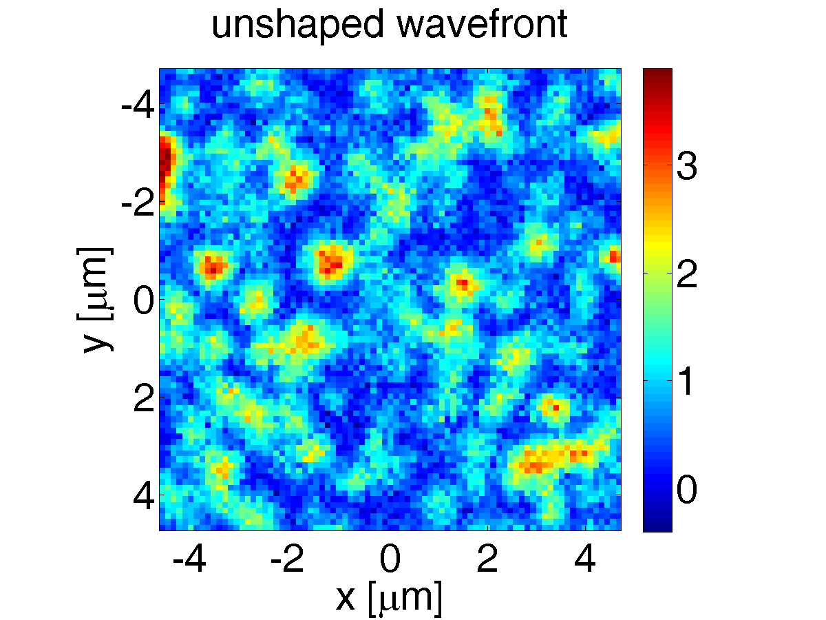
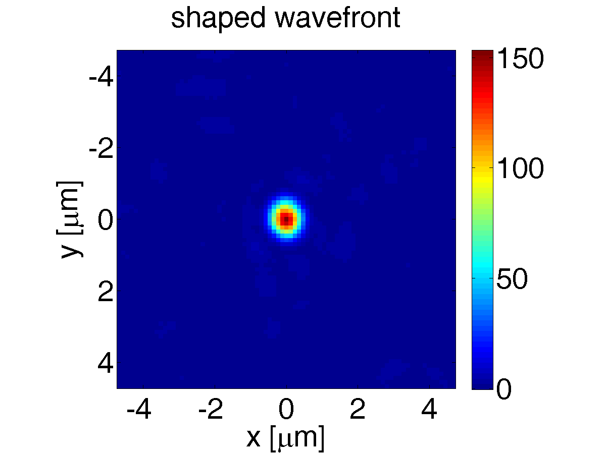
In the presence of many scattering events, a material becomes turbid and imaging is strongly hampered as increasingly little light is left over for direct imaging. By using phase shaping techniques, it is however possible to change the interference of the scattered light such that only constructive interference takes place at a specific place. This means that it is possible to create a focus behind a layer of turbid material, which we employ to create a novel type of microscope based on multiple scattering of light. The focus is illustrated in the two pictures below, where the intensity behind a turbid screen is shown once using a coherent light source (unshaped wavefront) and once with a shaped wavefront to create an interference generated focus. In both cases, the intensity is normalised to the average transmitted intensity passing through the turbid layer. this shows that in the focus, the intensity is increased by two orders of magnitude.
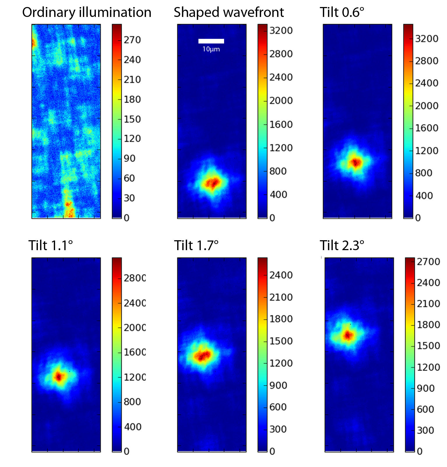
After creating a focus through a turbid layer, this focus needs to be scanned across a sample in order to be useful for microscopy. Using the optical memory effect, the interference pattern resulting in the focus is correlated over a certain angular range, making it possible to scan the focus over this angle by tilting the incoming illumination. This is shown in the figure below, for light focused through a piece of non-transparent tape.
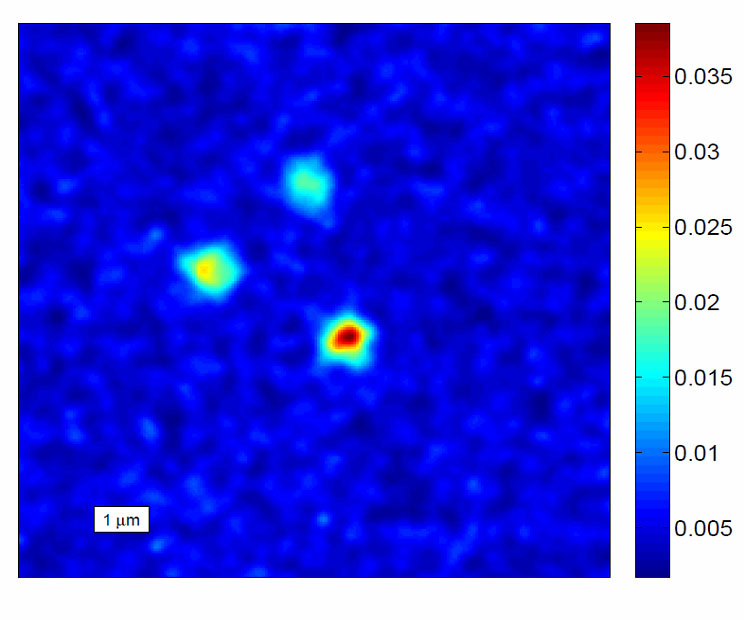
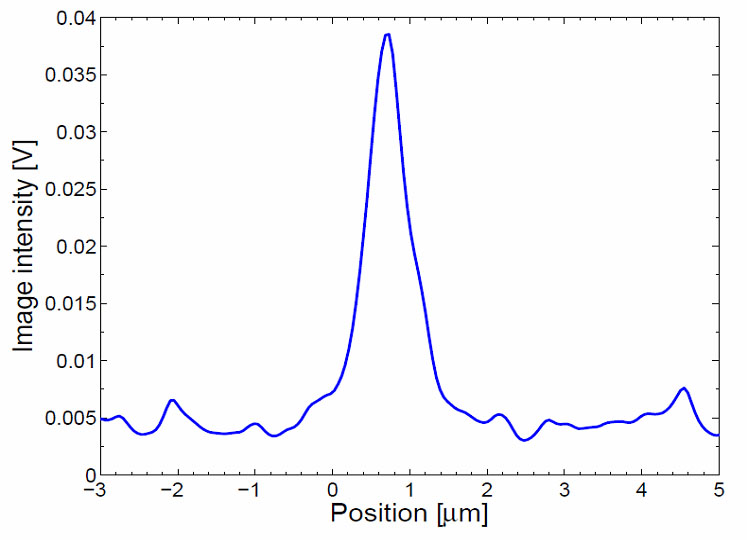
With such a scannable focus through a turbid layer, we have constructed a fluorescence microscope. Here, the focused laser beam is used to excite fluorescence in 200 nm sized bead. Only when the light is focused on the beads is the emitted fluorescence strong enough to be detected. Thus when the focus is scanned across the sample, an integrated measure of the fluorescence is sufficient to locate particles. This is shown in the figure below for three separate beads. The resolution can be gauged from a cross-section of the intensities across the beads and corresponds to 300 nm.
For more information consult the 2010 Optics Letter in the publications section.
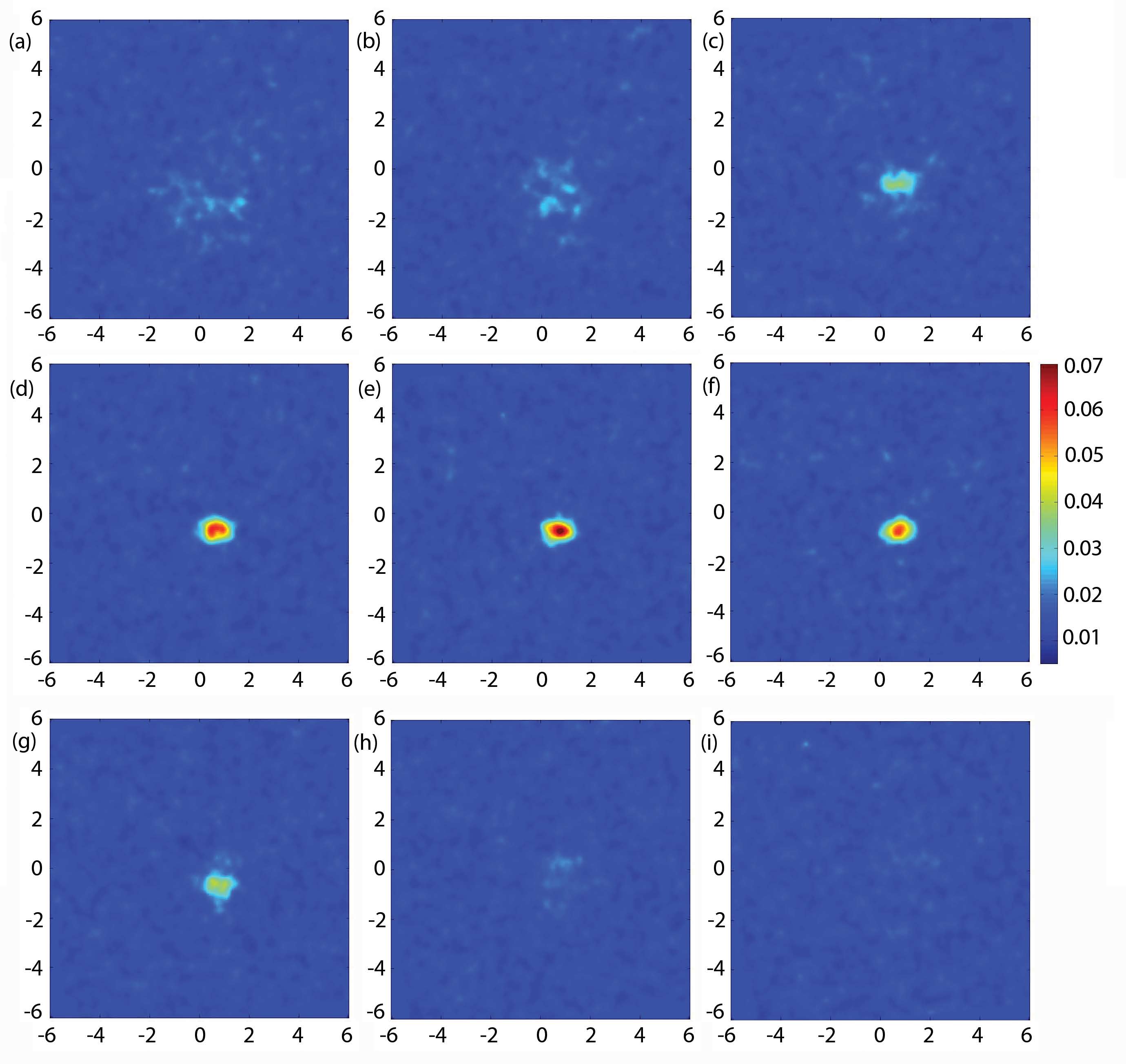
Using an additional parabolic pattern on the spatial light modulator, it is possible to also scan the wave-front shaping focus in the third dimension. This is shown in the figure below, where the cross-sections at different z-positions of a single fluorescent particle behind a turbid screen are shown. The turbid screen in this case is 15 mean free paths thick and te resolution is diffraction limited.For more details see our 2012 Optics Express publicaton.
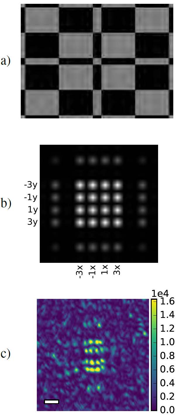
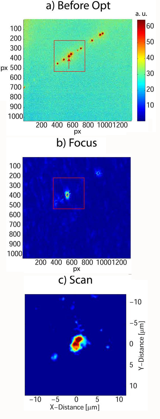
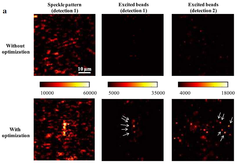
In order to be useful as a microscopy technique, the selective focusing has to be possible without optical access. For this purpose, the fluorescence of the structures of interest can be used as a contrast on which to optimise the wave front. Iteration of this scheme leads to the creation of a single focus on the brightest particle. the vicinity of that particle can then be scanned using the optical memory effect. The obtained focus and subsequent scan of a collection of fluorescent particles is shown in the figure below.
Single plane illumination microscopy in turbid media
- Given the geometry of illumination below, using an illumination and a detection objective, three dimensional imaging is possible at higher speeds with single plane illumination microscopy. Using wave-front shaping it is possible to obtain high quality light sheets for illumination even in the presence of turbid layers before the sample.
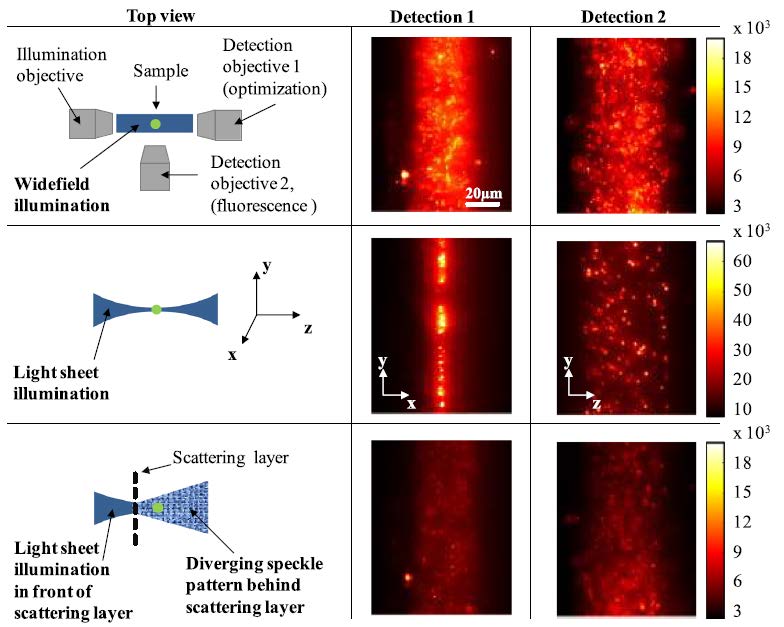
With such an optimised light sheet, high resolution imaging is therefore possible also in turbid environments, as shown below.
Structured illumination behind turbid media
After wave-front shaping, spatial light modulation can be used to change the illumination to one of a desired pattern by adding appropriate phase masks as shown below. This means that super-resolution microscopy is possible behind turbid layers.